Knee
Menisci
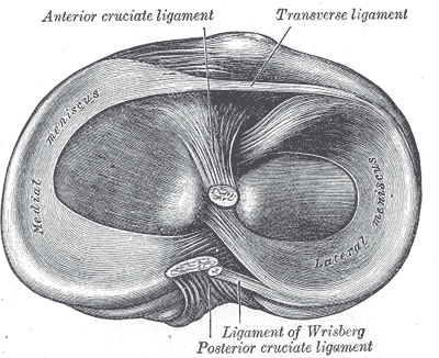
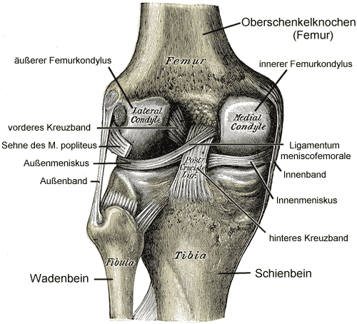
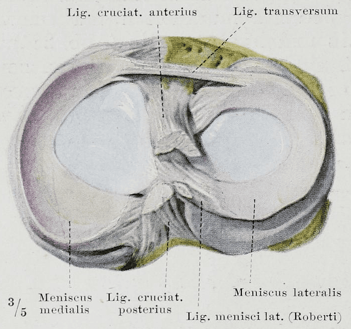
In each of our knee joints, there are two menisci. These are two large, fibrocartilaginous wedges. They differ slightly in shape, lie completely within the joint, and are enclosed by the joint capsule. The outer (lateral) meniscus has a more circular O-shape, while the inner (medial) meniscus has a more crescent-like C-shape. Both menisci are divided into an anterior and a posterior horn.
Role and Function
The role of the menisci is to compensate for the significant difference between the upper and lower leg bones of the knee joint and to reduce joint incongruity. This increases the stability of the knee joint and at the same time reduces the load on the hyaline articular cartilage surfaces of these bones.
Henry Vandyke Carter creator QS:P170,Q955620 derivative work: Hellerhoff Henry Graycreator QS:P170,Q40319 , Gray348-2, marked as public domain, details on Wikimedia Commons
Structure
The menisci are also a connective tissue structure composed of cells and matrix. Physiologically, they are comparable to articular cartilage (see also our blog post on Articular Cartilage). Menisci consist of approximately 75% water.
Both menisci are thickest in their outer region and become thinner toward their center. The thicker outer area also contains the most cells and has the richest nerve supply and blood flow.
Braus, Hermann, Braus 1921 284, marked as public domain, details on Wikimedia Commons
The alignment and orientation of the fibers vary greatly across the different regions depending on stress and physical demands.
Since the menisci only have a blood and nerve supply in their thicker outer zones, good quality synovial fluid is crucial for nourishing them (synovial fluid is produced by the joint capsule). To promote and support diffusion and osmosis processes, they need a regular alternation between loading and unloading to maintain their structure and function.
Henry Vandyke Carter Henry Gray, Gray348-de, marked as public domain, details on Wikimedia Commons
The menisci are fused with the joint capsule in their outer region, but the inner medial meniscus is attached much more firmly (due to the additional connection with the medial collateral ligament of the knee, which is more strongly connected to the joint capsule and the medial meniscus on the inner side than on the outer side). As a result, the outer lateral meniscus has more mobility than the inner medial meniscus—and because of this reduced mobility, the medial meniscus is correspondingly more susceptible to traumatic injuries.
Degeneration and Trauma
The most common causes of problems are, on the one hand, age-related or immobilization-induced degenerative changes, and traumatic injuries on the other.
When the joint is not moved, degenerative changes set in very quickly.
Immobilization leads to a very rapid loss of water and ground substance, which means the collagen network is no longer under tension (just like with articular cartilage), and the load-bearing capacity of the cartilage and menisci is severely reduced.
The resulting increased deformability of the menisci places greater strain on the fiber network. At the same time, the mobility of the menisci relative to the articular cartilage surface is also reduced.
In principle, any movements in your knee joint that involve a high range of motion and/or high velocity can cause injury to the menisci. Most often, trauma occurs through combined movements (flexion, inward buckling, and external rotation). As explained above, because the inner medial meniscus has less mobility, it is more vulnerable to injury and is therefore more frequently affected by traumatic injuries. It is not uncommon for such combined movements to also injure the medial collateral ligament (MCL) and the anterior cruciate ligament (ACL) (a condition known as the "unhappy triad").
Training
Just like with articular cartilage, exercise improves the micellar alignment in the menisci and thus enhances their tensile strength. Furthermore, reversible hypertrophy (more cartilage cells are produced) reduces incongruity by expanding the contact surfaces. This improves pressure elasticity and shock absorption in your knee joint.
Training and exercise don't just improve the adaptive capacity of cartilage tissue: strengthening your skeletal muscles and improving your coordination also relieves strain on the knee joint during movements—especially under higher loads—thereby protecting your tissue and body.
Therapy
To ensure optimal wound healing and regeneration of injured tissue, regular physiological loading is required. This is the only way your body can rebuild new, functional, and resilient tissue. Without appropriate physiological stimuli, inferior replacement or scar tissue of lower quality will develop.
When you have a knee injury, you should therefore keep moving and loading your knee in a reduced, modified way whenever possible.
In Physiotherapy and osteopathy at BodyLab, we know the anatomical and physiological factors inside out. When you face injuries or Conditions / issues, we know which therapeutic and training options are available, and we are happy to advise and guide you through them.
If active loading is not yet possible (after surgery or injury) or no longer possible, we can use passive joint techniques—sometimes under compression or traction—to improve the quality and function of your menisci as well as your entire joint as a functional unit, reducing pain and Conditions.
Once again: movement is life!
Whenever you need us, we are here for you!
Your BodyLab team, your specialists for movement and bodily Conditions
Osteopathy and Physiotherapy | Rehabilitation and Training
Zurich Altstetten
You can also find more information about cartilage tissue on our blog under Articular Cartilage.
Find out more in another blog post about our Intervertebral Disc - Structure & Function.
You can learn more about this on our page about meniscus tear.
Cover Image Credits
Henry Vandyke Carter Henry Gray, Gray349, marked as public domain, details on Wikimedia Commons