The Essentials
Articular cartilage - Hyaline cartilage
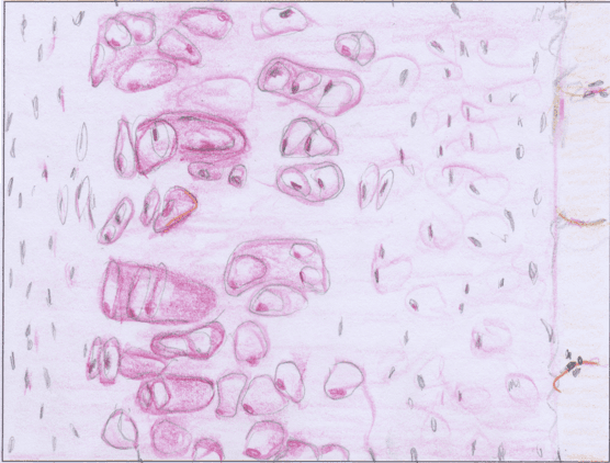
Characteristic of cartilage are the chondrocytes (cartilage cells), which lie within the cartilage ground substance (extracellular matrix, ECM) and produce it (as in all types of connective tissue). Depending on the type and amount of fibers in the ECM, three types of cartilage are distinguished: hyaline, elastic, and fibrocartilage.
In adults, all types of cartilage are largely free of blood vessels; their nourishment occurs through diffusion processes from the vascular perichondrium or, in the case of hyaline articular cartilage, directly through the synovial fluid (synovia) produced by the joint capsule.
Specific to all three types of cartilage is their high compressive elasticity, their viscoelastic deformability (bending stiffness), and their resistance to shear forces.
Hyaline articular cartilage
Function
Hyaline cartilage forms the protective layer covering the very sensitive and well-innervated bones in all synovial joints. It absorbs impact and compressive forces. By minimizing friction, it ensures smooth joint movement, which is crucially supported by the synovial fluid.
Structure and components
Its morphological structure consists of 4 different zones:

Zone I: Superficial cartilage zone (approx. 5%), main task: absorb shear forces and reduce friction
It is the thinnest of the 4 zones; water retention is highest; coated with synovial fluid; collagen fibrils are the thinnest and run parallel to the joint surface
Zone II: Middle cartilage zone or transitional zone (approx. 15%), main task: not specifically known
Collagen fibrils run obliquely to the surface and form arches (arcades)
Zone III: Deep or radial cartilage zone (approx. 40–60%), main task: absorption of compressive forces
Thickest layer; collagen fibrils run perpendicular to the joint surface; most active cartilage cells
Zone IV: Calcified cartilage zone (approx. 30%), main task: connecting articular cartilage to the bone
Relatively thin layer; the boundary between the deep and calcified cartilage zones is formed by the tidemark; collagen fibrils pass through the tidemark into the calcified zone and connect the softer, deformable cartilage with the hard, non-deformable articular cartilage; connect and stabilize the articular cartilage with the bone
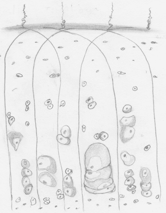
Due to its negative charge, the ground substance of hyaline cartilage can store a great deal of water (60–80%). The strong binding of water and collagen fibers creates a homogeneous network that can resist compressive loads extremely well—the primary task of hyaline articular cartilage.
The cartilage cells (chondrocytes) synthesize the necessary components of the ECM. However, synthesis activity decreases significantly after the growth phase is complete. It also depends on the quality and nutrient supply from the synovial fluid, the composition of the matrix, and the subchondral, well-vascularized bone. This poses a challenge, as cartilage has almost no blood vessels to supply nutrients. Nutrients reach the cells mainly through diffusion and osmosis. During compressive loading, water moves not only within the cartilage, but also penetrates into the synovial fluid and the subchondral bone. During the subsequent relief phase, the fluid flows from the underlying bone back into the cartilage, bringing with it the necessary nutrients required to maintain healthy articular cartilage.
Alternating pressure and relief promote the transport and exchange of these vital substances.
What does our articular cartilage need?
Healthy articular cartilage requires continuous synthesis and renewal of its fibrils and ground substance for its maintenance. The turnover time of hyaluronic acid is only 2–4 days, whereas that of fibrils is considerably longer. Keep in mind that the turnover time of collagen fibers is 300 to 500 days, which is a long time. Therefore, chronic underloading is the most harmful thing for the good function and high stability of our articular cartilage. And this is often an inevitable consequence of our modern sedentary lifestyle.
What effect does exercise have on cartilage tissue?
Training has a major influence on cartilage structure: the micellar arrangement (micelles: aggregated molecular complexes) is improved after training. This means an increase in tensile strength. Cartilage tissue responds to training stimuli with reversible hypertrophy (chondrocytes increase in number). Among other things, this reduces incongruence by enlarging contact surfaces, thereby improving compressive elasticity and shock absorption.
However, training and exercise do not only improve the adaptive capacity of cartilage tissue: stronger skeletal muscles and better technique/coordination also relieve joint movement under higher loads, protecting both the tissue and your body!
Osteoarthritis & Co.
Degenerative changes (wear and tear) are referred to as osteoarthritis. Contributing causes include reduced alternating cycles of loading and unloading, increasing ossification of cartilage tissue, and trauma.
Immobilization—due to a wide variety of causes—leads very quickly to severe demineralization with pathophysiological changes in our cartilage and bone tissue.
Therapy
The physiotherapists and osteopaths at BodyLab understand the anatomical and physiological factors and know what therapeutic options are available in case of injuries or Conditions / problems. We will gladly advise and instruct you on exercise options. If this is not yet possible (following surgery or injuries) or no longer possible, passive joint techniques—partially under compression or traction—can improve the quality and function of the articular cartilage as well as the entire joint as a functional unit, thereby reducing pain and Conditions.
Here too, the saying holds true once again: life means movement!
Whenever you need us, we are here for you!
Your BodyLab team, your point of contact for movement and Conditions of the body
Osteopathy and Physiotherapy | Rehabilitation and Training
Zurich Altstetten
Learn more about our menisci in another blog post.
Find out more on our page about knee osteoarthritis.