Knee
ACL injury

A cruciate ligament injury is incredibly common – especially in sports and recreational activities – due to the high stress combined with the great mobility of the knee.
Because it is so common, this topic is widely discussed and thoroughly researched in the scientific community.
In the last "Sport Schweiz" survey conducted in 2014 by the Sport and Physical Activity Observatory (Federal Office of Sport FOSPO), aiming to paint a detailed picture of the current sporting activities of the Swiss population, 35.4% of respondents stated that they ski [1]. Along with hiking (44.3%), cycling (38.3%), and swimming (35.8%), skiing is one of the most popular sports among Swiss people [1].
Compared to hiking, however, the risk of injury to the knee and the cruciate ligaments, specifically the anterior cruciate ligament (ACL), is significantly higher when skiing. Data from the latest survey by the Swiss Council for Accident Prevention (bfu.ch) showed that an average of 22,260 people are injured each year while hiking, compared to approximately 51,700 while skiing [2] – 30.9% of these injuries affect the knee joint [3], primarily the anterior cruciate ligament. We see this firsthand at BodyLab, Osteopathy and Physiotherapy, Rehabilitation and Training in Zurich every single winter.
Outside of the ski season, football is one of the most popular recreational sports in the world, and Switzerland is no exception. Studies show that this sport also frequently results in lower extremity injuries, particularly to the knee [4].
Various studies estimate that up to approximately 17.6% of football-related injuries presenting to the emergency department affect the knee [5,6,7,8,9].
The majority of these football injuries resulted from non-contact mechanisms (85% of injuries without opponent involvement), although contact injuries during tackles are of course also possible [10,11].
Anatomy and Function of the ACL
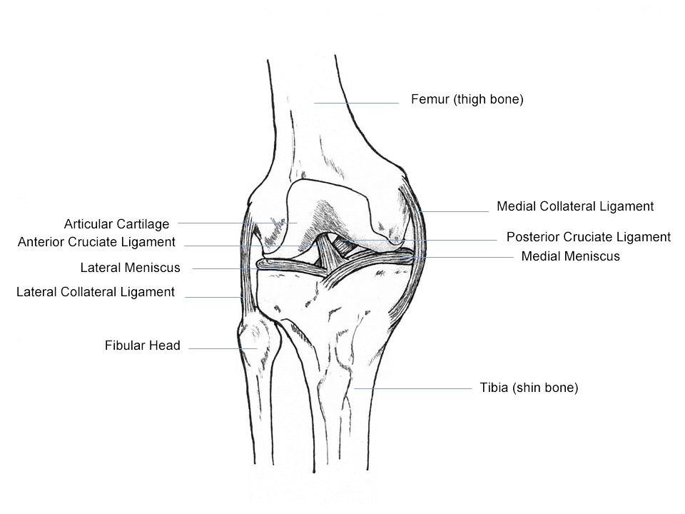
The ACL is one of the most critical ligament structures around and inside the knee joint. Together with the posterior cruciate ligament (PCL) and the collateral ligaments, it helps stabilize the knee joint by stretching from the posterior end of the femur to the anterior part of the tibial head.
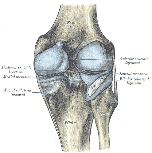
The tight connective tissue of the ACL consists mainly of type I collagen fibers and is divided by type III fibers into two primary bundles with different functions. The anteromedial portion is about 36mm long on average and becomes tense from 30° of knee flexion. The main task of the anteromedial bundle is to stabilize against anterior tibial translation, meaning it prevents the shinbone (tibia) from sliding forward.
The posterolateral bundle is approximately 18mm long and becomes tense at less than 30° of flexion. This part of the ACL, together with other ligament structures, serves to stabilize rotational forces in the knee joint.
The ACL is not just a passively stabilizing structure in the knee joint. Thanks to Pacinian and Ruffini corpuscles, as well as other free nerve endings in the insertion area of the ligament, vibration sensations, stretching, and shearing forces are perceived. This information is used for joint position awareness to generate adapted muscular responses. In the case of the ACL, this means an increased tensioning of the hamstrings to reduce anterior tibial translation.
Mechanisms of Injury
A cruciate ligament injury can be triggered by both contact and non-contact mechanisms.
Typical mechanisms of injury – and the most common way to suffer an anterior cruciate ligament injury – involve a combination of twisting and buckling trauma of the knee joint under load (strong valgus buckling of the knee (inward) during slight flexion of the knee combined with rotation).
Mechanisms of Injury in Football
Due to studs fixing the foot to the grassy ground, massive uncontrolled loads can act on the knee. As a result, 85% of all cruciate ligament injuries in football occur in non-contact situations [10,11]. Video analyses of professional football players also show an increased risk of ACL injury through non-contact situations within the first 9 minutes of the match, and that the risk of injury is greater during defending and tackling activities [12,13,14].
Mechanisms of Injury in Skiing
Using video analysis of elite alpine skiers after an ACL rupture, Bere et al. identified three accident mechanisms [15]:
Slip-Catch
Dynamic Snowplough
Landing after jumps
Slip Catch
In nearly 50% of all cases, a so-called slip-catch mechanism was the cause. Here, the skier lost snow contact with the outer ski during a turn. Upon regaining snow contact, the ski caught, leading to increased knee flexion, internal rotation of the tibia, and inward buckling of the knee (valgus position). This mechanism often brings along an injury to the medial meniscus and the medial collateral ligament – the so-called Unhappy Triad.
Dynamic Snowplough
Another accident scenario (15%) was the dynamic snowplough. Here, the skier was thrown off balance while leaning back, causing the less weighted leg to drift away and forcing the skier into a split position. The ski rolled from the outer edge to the inner edge, caught in the snow, and forced the knee back into a valgus position and tibial internal rotation.
Landing after Jumps
The final mechanism (20%) occurred when landing after jumps. The skier lost balance, pushing the body weight backward. In trying to regain balance, a combination of increased compression in the knee and a major anterior tibial shift (forward movement of the tibia relative to the femur) subjected the ACL to extreme loads.
Considering that professional skiers deal with much greater forces due to speed and equipment, these mechanisms cannot be fully applied to recreational athletes. However, the individual movement factors (strong flexion, valgus position, and tibial internal rotation) remain identical.
For recreational athletes, so-called forward twisting falls are the most common accident mechanism at 54% [16]. This involves a valgus position and internal tibial rotation after catching the ski edge while turning.
The second most common accident scenario is called a backward twisting fall or phantom foot [6]. This rupture mechanism occurs in 26% of all cases. Here, the center of gravity is shifted backward and downward onto one leg by dropping the pelvis (increased knee flexion), whether while turning or after a fall, and the weight presses onto the tail of the ski. Combined with stiff boots, the tibia is forced forward, and the ACL ruptures.
Knee trauma often leads to accompanying injuries (see Unhappy Triad, medial collateral ligament injuries, meniscus injuries, cartilage damage, secondary osteoarthritis).
Contributing Factors / Equipment
The advent of carving skis showed that the increased aggressiveness of the ski due to its sidecut, combined with stiff ski boots, leads to more ACL injuries [15]. Sharper turns can be made, and more speed is generated. This requires more strength from the skier and makes controlling the equipment harder. Due to the stiffness of the ski boots, when shifting weight backward, the tibia is pushed anteriorly, placing more stress on the ACL. Furthermore, video analyses revealed that in 100% of all slip-catch cases, the binding did not release or released too late [15].
Biomechanics in the Knee after ACL Rupture
First and foremost, there is a combination of anterior knee instability with anterior tibial translation and rotational instability. This impacts the entire biomechanics of the knee. The center of rotation in the knee joint shifts medially (inward). This leads to increased stress on the collateral ligaments (medial and lateral collateral ligaments) and the horns of the meniscus, which also puts more load on the cartilage surfaces. For example, the medial collateral ligament is stressed up to 140% more, while the posterior outer structures have to perform up to 400% more work [17]. During walking, this shows up as prolonged flexion during the stance phase and increased anterior tibial translation towards the end of the stance phase [17].
Surgery or Conservative Treatment
After an anterior cruciate ligament injury, you basically have two options – surgical reconstruction of the ACL followed by physiotherapy, or conservative treatment with physiotherapeutic measures – with the option of surgery if needed.
In Switzerland, the Swiss Medical Board addressed this issue (and also published an information sheet [19]): After an extensive literature review, the panel concluded that both surgical and conservative approaches can achieve satisfying results – allowing individuals to return to their previous physical activities in most cases. This is shown by studies that evaluated outcomes at one, five, and up to 15 years after a cruciate ligament injury. No significant differences were found when comparing conservative treatment with surgery [18].
With conservative treatment, if knee instability persists during the course of therapy, surgery can still be performed later without losing any valuable time.
Conclusion: "When choosing a treatment method, the respective advantages and disadvantages (namely the surgical risk) must be carefully weighed; if in doubt, get a second opinion." [19]
Conservative Therapy
The goal of conservative therapy and physiotherapy after a cruciate ligament injury is to restore physiological mobility, as well as the stability of the knee joint and the leg axis. Following injuries, the body responds with specific biological processes (inflammatory phase, proliferation phase, remodeling phase) that a physiotherapist must consider to facilitate the fastest possible healing and restoration of function.
Corresponding to the healing phases, short-, medium-, and long-term goals emerge for therapists and patients. In the first 1-2 weeks (inflammatory phase), reducing swelling and pain relief through passive measures such as lymphatic drainage and low-load/load-free movements are key focus areas of the treatment [20].
During the proliferation phase (weeks 3-6), the focus is on regaining mobility, progressive loading (possibly weaning off crutches, moving from partial to full weight-bearing), improving stability, and strengthening muscular deficits in closed kinetic chains. This is achieved through both passive and active exercises [20].
From the 7th post-traumatic week (remodeling phase), the complete restoration of leg axis stability (pelvis-leg axis) and building up musculature with strength training exercises using weights in the gym are central. The ultimate goal is to be pain-free in everyday life and to make a successful return to your previous sporting activities [20].
Whenever you need us, we are here for you!
Your BodyLab Team, by your side back to daily life and sports
Osteopathy and Physiotherapy | Rehabilitation and Training
Zurich Altstetten
References
[1] Sporting activity and sporting interest of the Swiss population.
Lamprecht, M., Fischer, A. & Stamm, H.P.
Sport Schweiz 2014: Sporting activity and sporting interest of the Swiss population. Magglingen: Federal Office of Sport FOSPO.
[2] Injured persons by sport and age, Ø 2010–2014
[3] Injury location among UVG-insured persons by sport (per 100 injured), Ø 2008–2012
Woods C, Hawkins R, Hulse M, Hodson A.
Br J Sports Med. 2002;36(6):436-441. doi:10.1136/bjsm.36.6.436.
[5] The FIFA 11+ injury prevention program for soccer players: a systematic review.
Sadigursky D, Braid JA, De Lira DNL, Machado BAB, Carneiro RJF, Colavolpe PO.
BMC Sports Sci Med Rehabil. 2017;9:18. doi:10.1186/s13102-017-0083-z.
Volpi P, Bisciotti GN, Chamari K, Cena E, Carimati G, Bragazzi NL.
Muscles Ligaments Tendons J. 2016;6(4):480-485. doi:10.11138/mltj/2016.6.4.480.
[7] Soccer-related injuries treated in emergency departments: 1990-2014.
Smith NA, Chounthirath T, Xiang H.
Pediatrics. 2016;138(4). doi:10.1542/peds.2016-0346.
Leininger RE, Knox CL, Comstock RD.
Am J Sports Med. 2007;35(2):288-293. doi:10.1177/0363546506294060
[9] Childhood soccer injuries treated in U.S. emergency departments.
Adams AL, Schiff MA.
Acad Emerg Med. 2006;13(5):571-574. doi:10.1197/j.aem.2005.12.015.
Woods C, Hawkins R, Hulse M, Hodson A.
Br J Sports Med. 2002;36(6):436-441. doi:10.1136/bjsm.36.6.436
[11] Severe injuries in football players. Influencing factors.
Chomiak J, Junge A, Peterson L, Dvorak J.
Am J Sports Med. 2000;28(5 Suppl):S58-68. doi:10.1177/28.suppl_5.s-58.
[12] Anterior cruciate ligament injury in elite football: a prospective three-cohort study.
Waldén M, Hägglund M, Magnusson H, Ekstrand J.
Knee Surg Sports Traumatol Arthrosc. 2011;19(1):11-19. doi:10.1007/s00167-010-1170-9.
Waldén M, Krosshaug T, Bjørneboe J, Andersen TE, Faul O, Hägglund M.
Br J Sports Med. 2015;49(22):1452-1460. doi:10.1136/bjsports-2014-094573.
Grassi A, Smiley SP, Roberti di Sarsina T, et al.
Eur J Orthop Surg Traumatol. 2017;27(7):967-981. doi:10.1007/s00590-017-1905-0.
BereT, FlørenesTW, KrosshaugT, et al.
Am J Sports Med. 2011;39:1421–1429
Ruedl G, Webhofer M, Linortner I, et al.
Int J Sports Med. 2011; 32 (10):801-6.
[17] Rupture of the anterior cruciate ligament. Biomechanical effects on the knee joint.
PD Dr. M. Herbort, Prof. Dr. C. Fink:
in: Arthroskopie (01/2015)
[18] Review technical report "Rupture of the ACL: surgical or conservative treatment?"
[20] ACL Rupture Therapy, AGA Knee Ligament Committee
Cover Image Credit
Kari Stammen, Human Knee Anatomy, marked as public domain, details on Wikimedia Commons