The Essentials
Nerve cells (Neurons)
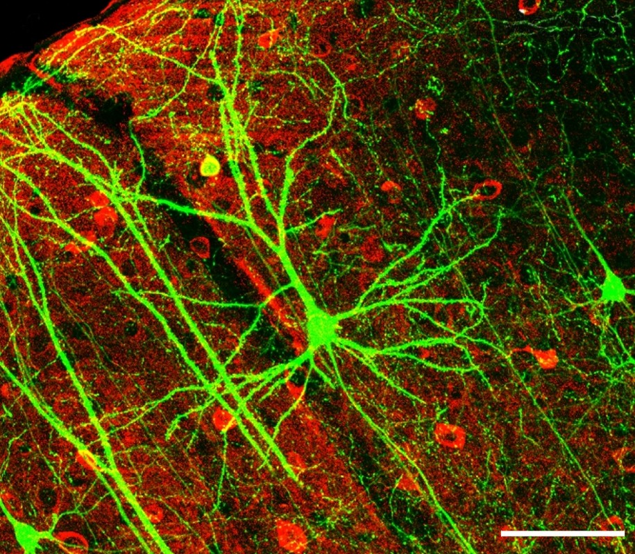
A neuron (nerve cell) is the structural and functional unit of our nervous system. Billions of these neurons form a functional mosaic through countless connections between cells, enabling the perception and interpretation of sensations (sensory stimuli), storage of experiences (memory), and reactions (behavior).
The nervous system is divided into a central nervous system (CNS, voluntary, conscious, or somatic nervous system) and an autonomic nervous system (ANS, involuntary nervous system). The CNS includes all anatomical structures surrounded by our meninges.
The CNS controls conscious, targeted processes, such as active movements via the skeletal muscles. The ANS controls unconscious (autonomous) activities, such as the digestive system, blood pressure, and much more.
Together, the CNS and ANS form two systems for controlling the human body. In addition, the endocrine system (hormone system) represents another instrument for transmitting information and control, which is regulated by and often considered part of the autonomic nervous system.
All of these systems share the same goal: transmitting information. They ensure that tissues and organs can communicate seamlessly with one another!
The difference is that the nervous system (NS) transmits signals electrically via neurons. This is relatively fast and can be easily switched on and off.
In contrast, the hormonal system relies on chemical signal transmission through the bloodstream, which is slower and cannot be switched on and off as quickly or precisely as the NS.
Structure of a Nerve Cell
Nerve cells can generate, transmit, and – very importantly – modify stimuli (such as pain) within the cell network!
A neuron consists of a cell body (soma) which has many projections of the nerve membrane. These projections form dendrites (there are countless of these, similar to branches on a tree) that receive (afferent) signals from other (nerve) cells. There is also an axon (which can partially form collaterals). This is a relatively long conduit, ranging from a few millimeters up to 1.5 meters (note: in giraffes or large mammals, these can obviously be even longer!). The electrical signal is transmitted along the axon to the end terminal of the nerve cell, known as the synapse. Synapses act as the contact points with other cells, transmitting the signal to the dendrites of the next nerve cell, to skeletal muscle cells, gland cells, or organs, triggering an action.
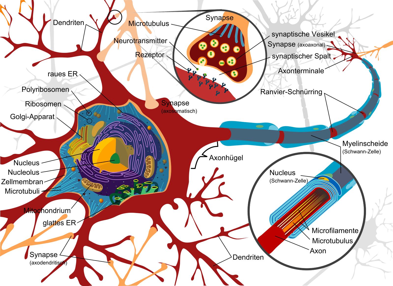
Nerve cells Complete neuron cell diagram de, marked as public domain, details on Wikimedia Commons
Nerve cells (along with liver cells) are the metabolically most active cells in the body. The neuron is also a highly flexible unit and is capable of diverse growth and transformation processes, especially in the area of the dendrites. It can be trained!
The axon is surrounded by connective tissue cells (known as glial cells: Schwann cells in the periphery, or astrocytes and oligodendrocytes in the CNS). These provide structure and support to the axon and form a more or less thick insulating layer (myelin sheath) that influences conduction speed and makes electrical signal transmission possible in the first place. Together with the axon, this sheath forms the nerve fiber.
Because of this insulating layer, the CNS actually consists of 90% connective tissue and only 10% actual nerve cells!
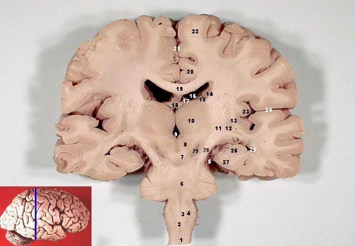
John A Beal, PhD Dep’t. of Cellular Biology & Anatomy, Louisiana State University Health Sciences Center Shreveport, Human brain frontal (coronal) section description 2, CC BY 2.5
The white color comes from the myelin, due to the accumulation of myelinated axons; the gray color represents the unmyelinated axons or the nerve cell bodies! That is why the cerebral cortex is gray, as it contains a particularly high concentration of cell bodies. For the same reason, a gray butterfly shape is visible inside the spinal cord (where the cell bodies are located), surrounded by white matter (the pathways, i.e., axons).
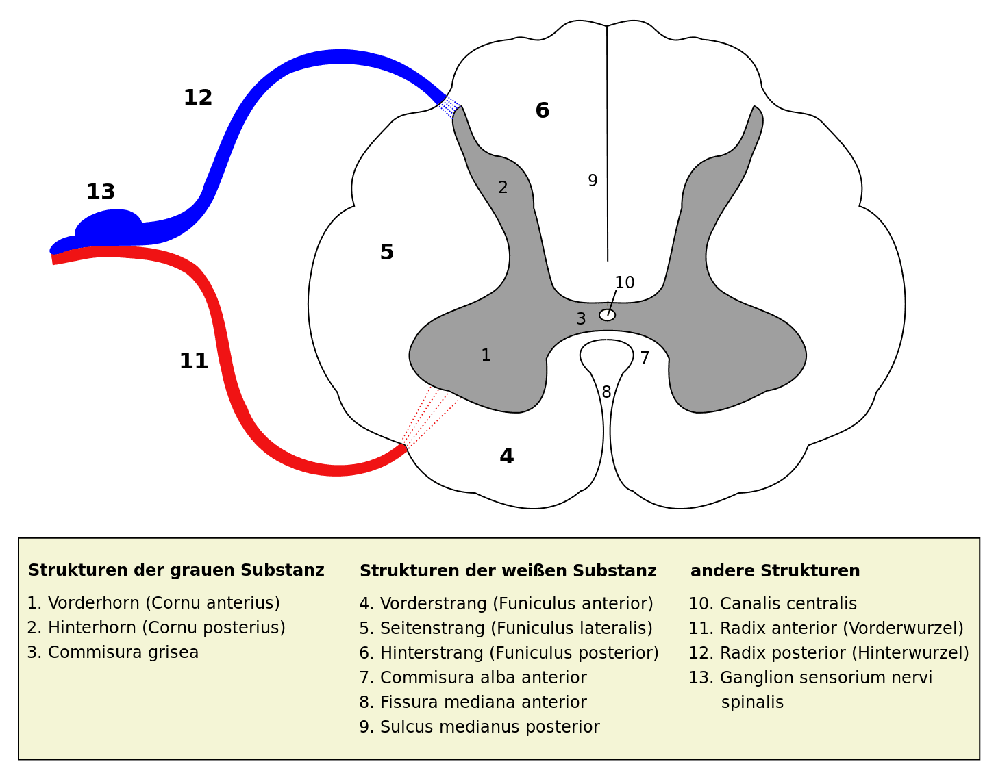
Spinal cord cross section User:Polarlys, Medulla spinalis – Querschnitt – German and Latin, CC BY-SA 3.0
Motor Neurons (Afferent Signals): A Movement
Simply put, the cell bodies are located in the brain (1st motor neuron) and in the spinal cord (2nd motor neuron). To execute a movement, the signal travels from the brain via descending pathways to the spinal cord, where it is switched (in the anterior horn) to another motor neuron and transmitted directly from there to the skeletal muscles.
Executing a voluntary movement therefore requires 2 nerve cells, with the signal being switched once.
Sensory Neurons (Efferent Signals): A Sensation
Sensory sensations are conducted from free nerve endings or receptors via the axon to the posterior horn of the spinal cord. However, the cell body of this 1st sensory neuron lies outside the spinal cord, specifically in the spinal ganglia. In the spinal cord, initial processing occurs via interneurons to a second sensory neuron, which ascends toward the brain. Before reaching the cerebrum, the signal passes through the thalamus, where it is switched to a 3rd sensory neuron.
Sensory signals are thus conducted to the brain through at least 3 neurons, requiring them to be switched twice. This allows efferent signals to be modulated, modified, and distributed in a much more versatile way.
As we will see in a later blog post, this opens up exciting therapeutic possibilities for us!
Diseases, challenges, and treatment options will also be covered in a future blog post. Stay tuned.
If you need us, we are here to support you!
Your BodyLab Team, your specialists for physical complaints
Osteopathy and Physiotherapy | Rehabilitation and Training
Zurich Altstetten
Title Image Credits
Microscopic image of the cerebral cortex of a mouse. Wei-Chung Allen Lee, Hayden Huang, Guoping Feng, Joshua R. Sanes, Emery N. Brown, Peter T. So, Elly Nedivi PLoSBiol4.e126.Fig6fNeuron, CC BY 2.5