The Essentials
Joint capsule and ligaments
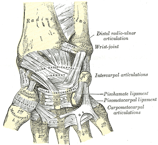
The joint capsule (Capsula articularis) and the ligaments surround every true joint in the body. The capsule encloses and forms the joint space, producing the joint fluid (synovial fluid). The movements of a joint are regulated and guided by the capsule and, even more so, by the ligaments. They directly influence the passive stability of a joint.
Function
A primary task of the capsule is producing high-quality, sufficient synovial fluid. This joint fluid enables smooth, friction-free movement between the joint partners. Furthermore, the synovia is crucial for nourishing the joint cartilage and the menisci, providing them with essential nutrients. The capsule also transmits vital information regarding the position and movement of the joint (proprioception). This is why it is richly innervated and equipped with numerous receptors. Most muscles are fused with the joint capsule; they tension it to prevent it from getting pinched between the moving bones. The ligaments function to control and limit the range of motion, sharing responsibility for the (passive) stability of the joints. They are also packed with numerous receptors, though these only respond later and under higher loads.
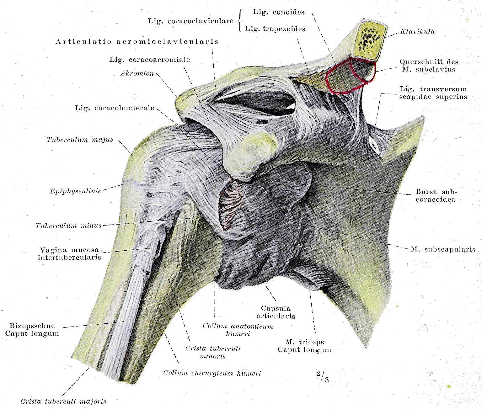
Braus, Hermann, Braus 1921 147, marked as public domain, details on Wikimedia Commons
Structure
The capsule is composed of two layers: an outer, more robust and firmer layer (membrana fibrosa) that protects and supports the delicate inner membrane.
The inner, more delicate layer—the so-called membrana synovialis (which can be further subdivided)—forms numerous folds. This is the body's natural way of increasing an (active) surface area (similar to the convolutions of the brain). The innermost layer of the membrana synovialis connects with the tangential layer of the joint cartilage, while the outer layer of the membrana synovialis connects with the intracapsular periosteum (the bone membrane inside the joint capsule). See also our blog post on Joint Cartilage.
In areas of the membrana fibrosa that are regularly subjected to specific mechanical loads and forces, the path of the collagen fibers adapts accordingly: the fibers align with the direction of the force acting upon them (much like bone trabeculae). Consequently, the capsule often becomes thicker in these areas, and we refer to them as ligaments. These are nothing less than an adaptation of the membrana fibrosa to mechanical stress and form an integral part of the joint capsule. These ligaments are typically flat and band-shaped.
There are also some ligaments that do not have a direct connection to the capsule (e.g., the lateral collateral ligament of the knee). However, even these are always connected to the capsule via loose connective tissue. These ligaments usually have a significantly rounder and more tendon-like structure.
The intracapsular ligaments represent a special feature; they lie inside the joint capsule and have no connection to the membrana fibrosa, but are connected to the membrana synovialis (e.g., the cruciate ligaments).
The blood supply to the ligaments is lower than that of the capsule. Therapeutically, this means that intracapsular ligaments heal and regenerate better and faster than extracapsular ligaments, which lack a direct connection to the capsule.
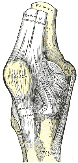
Anterior knee joint capsule, Henry Vandyke Carter Henry Gray, Gray345, marked as public domain, details on Wikimedia Commons
Degeneration
During the aging process, and even more so after injuries, there is an increase in Type II collagen, which grows into the capsule starting from the capsule-bone junction. This leads to chondrification of the capsule (fibrous cartilage).
The tissue becomes tighter and less elastic, and the number of so-called soluble cross-links decreases, while the number of non-soluble cross-links increases significantly. This shift is also observed in diabetes mellitus.
Overall, the water content of the capsule and ligaments decreases with age, causing further loss and decline of elasticity and, as a direct consequence, load capacity in older age.
Immobilization also accelerates these changes; in fact, they occur to an even greater extent. As a result, the length of an immobilized ligament increases more in relation to the load applied to it, reducing the stability of the affected joint. The capsule shrinks due to changes in the membrana fibrosa, which decreases mobility and flexibility.
Interestingly, the body has built-in protective mechanisms to ensure we rarely reach the maximum load limits of our connective tissue structures. Studies show that during physiological daily movements of the knee joint, we only use about 10% of the maximum load capacity of the anterior cruciate ligament and only about 30% of the patellar ligament.
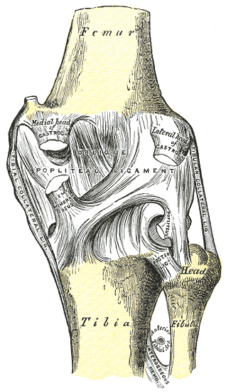
Posterior knee joint capsule, Henry Vandyke Carter Henry Gray, Gray346, marked as public domain, details on Wikimedia Commons
What our joint capsule needs
When a joint moves—and with it, the capsule—oxygen and nutrients travel from the extracapsular vessels to the intracapsular vessels of the membrana synovialis. They then reach the synovial fluid via diffusion and osmosis. Through movement and the constant alternation between tension and relaxation, nutrients are transported from the vessels into the interior of the joint and back. Ligaments require loading stimuli, just like those generated with every movement. Without these—for example, after prolonged immobilization—the load-bearing capacity of a ligament decreases rapidly. Whether the load capacity of a healthy ligament can be improved at all remains unresolved.
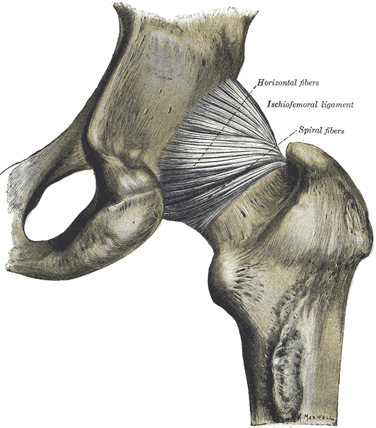
Hip joint capsule ligaments, Henry Vandyke Carter Henry Gray, Gray340, marked as public domain
Training
Whether and to what extent training has a direct impact on the quality of a healthy capsule and its ligaments is debated and questionable. Research has shown that training has only a minor influence on the thickness of ligaments and thus their stability. What is undisputed, however, is the devastating effect of immobilization, which leads to massively reduced load capacity and a decline in ligament quality!
But as we know from our previous blogs, movement with alternating loading and unloading promotes various positive effects: circulation increases, synovial fluid production is stimulated, and the exchange of substances is enhanced and improved. This all contributes to enhancing the quality of the functional unit. By improving technique and coordination, proprioception is also boosted alongside the neural system.
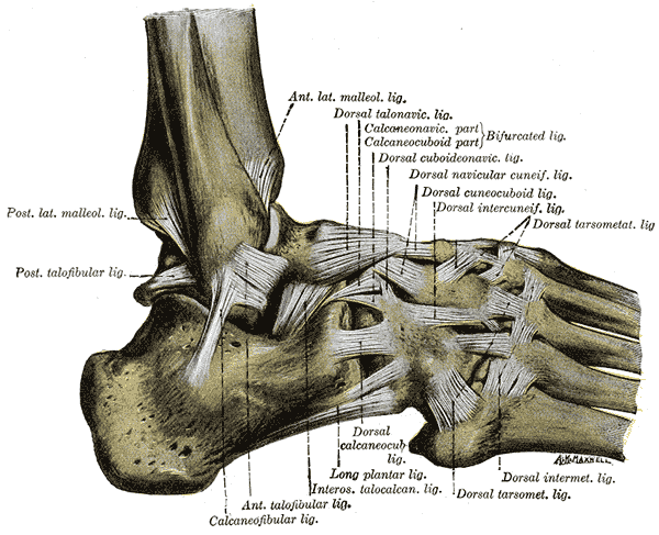
Henry Vandyke Carter creator QS:P170,Q955620 Henry Gray creator QS:P170,Q40319, Gray355, marked as public domain, details on Wikimedia Commons
Therapy
It is assumed that stimulating the capsule receptors reflexively leads to pain relief in the central nervous system and a reduction in sympathetic nervous activity. This normalizes physiological processes. Additionally, the mobility of the treated joint is improved. Both manual therapy and active exercise forms stimulate these joint receptors.
Furthermore, recent studies show that the formation of scar tissue is linked to a lack of physiological loading stimuli during the healing phases. It is crucial to monitor loading stimuli throughout rehabilitation to prevent new ruptures, as the load capacity of the capsule and ligaments, as well as their proprioception, is significantly reduced!
The physiotherapists and osteopaths at BodyLab are experts in anatomical and physiological conditions. In the event of injury or discomfort, they know exactly which therapeutic options are available. We are happy to advise and guide you on training and exercise options. If active exercise is not yet possible (following surgery or injury) or no longer possible, passive joint techniques—sometimes using compression or traction—can improve the quality and function of the joint cartilage and the entire joint as a functional unit, thereby reducing pain and discomfort.
Once again, the rule is: life is movement!
Whenever you need us, we are here for you!
Your BodyLab Team, your specialists for movement and body aches
Osteopathy and Physiotherapy | Rehabilitation and Training
Zurich Altstetten
Cover Image Credits
Henry Vandyke Carter Henry Gray, Gray334, marked as public domain, details on Wikimedia Commons