Shoulder
Shoulder Impingement - Subacromial Impingement Syndrome

Five different joints contribute to the movements of the shoulder girdle – the glenohumeral joint, the acromioclavicular joint, the sternoclavicular joint, the subacromial gliding mechanism, and the scapulathoracic gliding space. Because of this anatomical design, the shoulder girdle possesses a massive range of motion – however, this also makes it more susceptible to injuries and shoulder pain. To ensure a flawless biomechanical movement sequence, the thoracic spine must also be fully mobile, in addition to the mechanics of the five shoulder joints.
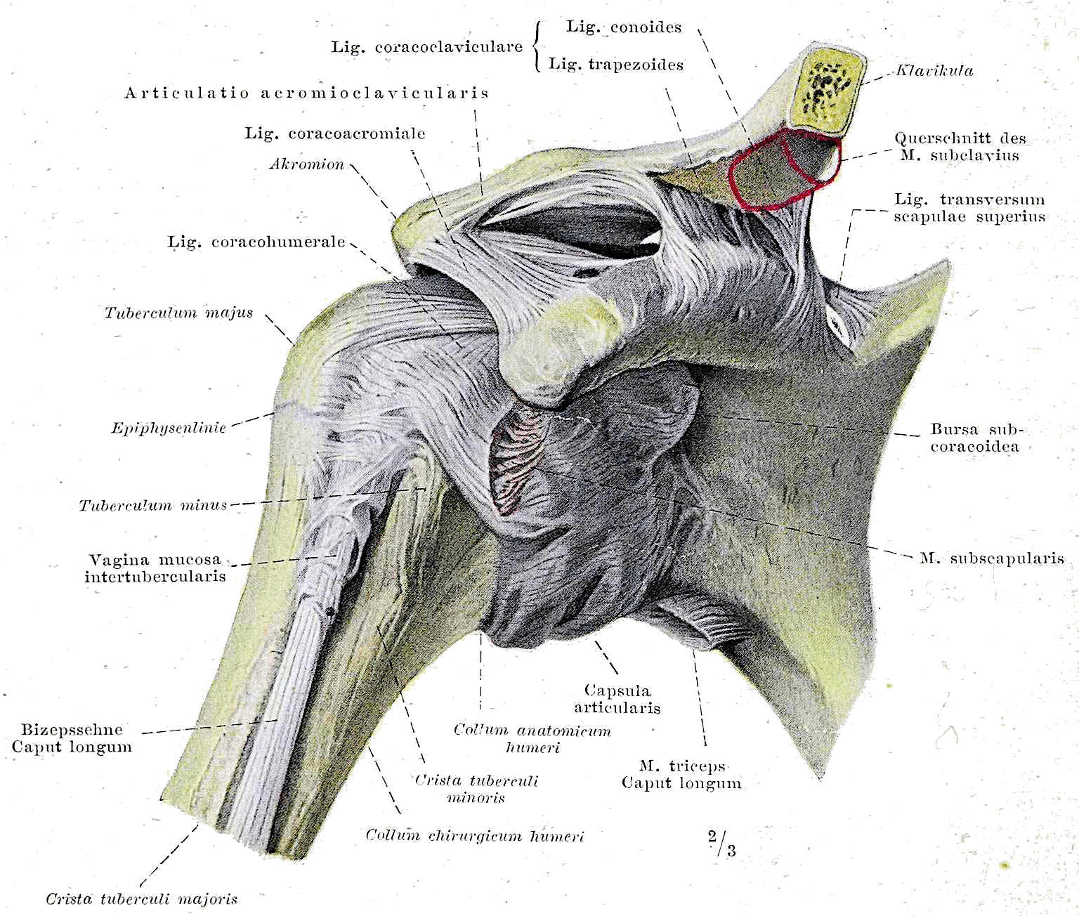
Braus, Hermann, Braus 1921 148, marked as public domain, details on Wikimedia Commons
The Anatomical Structure of the Shoulder Joint
The glenohumeral joint (commonly known as the shoulder joint) consists of the head of the humerus (upper arm) and the glenoid cavity (socket) on the shoulder blade. As a classic ball-and-socket joint, the glenohumeral joint is key to the shoulder girdle's impressive range of motion.
The surface of the humeral head is three to four times larger than that of the glenoid cavity. While this enables incredible shoulder mobility, it comes at the cost of stability. The fibrocartilaginous glenoid labrum on the edge of the socket is designed to compensate for this incongruence alongside the entire capsulo-ligamentous apparatus and boost the stability of the shoulder joint. However, that stability is primarily secured by the powerful shoulder musculature.
The acromion and the clavicle (collarbone) form the acromioclavicular joint. When moving the arm above shoulder height, the clavicle rotates around its own axis. Ligaments (acromioclavicular, coracoacromial, and coracoclavicular) keep the acromioclavicular joint in position during the longitudinal and craniocaudal rotation of the clavicle.
The same applies to the sternoclavicular joint – formed by the clavicle and the sternum (breastbone) – at the other end of the collarbone. The sternoclavicular joint also features an articular disc (similar to an intervertebral disc) to balance out the misalignment between the joint surfaces of the clavicle and the sternum.
After keeping the arm in a protective posture for too long, adhesions can form in the capsulo-ligamentous apparatus – this affects the ligaments of the acromioclavicular, sternoclavicular, and glenohumeral joints. This restricts mobility and disrupts the biomechanical flow. In the worst cases, adhesion or shrinkage of the capsule over time, combined with complications, can lead to a frozen shoulder with severe limitations on movement.
The scapulathoracic gliding space and the subacromial gliding mechanism are not true joints, yet they play a critical role in shoulder biomechanics. The scapulathoracic gliding space describes the movement of the shoulder blade (scapula) on the rib cage. The subscapularis and serratus anterior muscles lie in between. In shoulder problems, it is common to see that the scapula cannot be sufficiently stabilized against the chest wall, causing it to move prematurely during arm motions.
The subacromial gliding mechanism is the space between the acromion and the head of the humerus, containing two bursae between the acromion and the rotator cuff. The rotator cuff – consisting of the infraspinatus, supraspinatus, subscapularis, and teres minor muscles – plays a central role. Its main job is to center the humeral head within the glenoid cavity. If one of these muscles is too weak, overloaded after an injury, or overused from repetitive movements, this centering and the biomechanical flow of the shoulder are compromised. Calcifications or thickening of the supraspinatus tendon, which runs through the subacromial gliding mechanism, cause a similar issue.
The supraspinatus muscle runs underneath the acromion, from the supraspinous fossa of the scapula to the head of the humerus. If the function of the supraspinatus is impaired, the initial phase of lifting the arm sideways (abduction) is disrupted. More importantly, due to the external rotation function of the supraspinatus, it centers the humeral head posteriorly in the glenoid cavity. Without this centering, lifting the arm sideways causes a pinch in the subacromial space – the humeral head glides upward too quickly and the greater tubercle on the humerus does not rotate backward in time, hitting the acromion and pinching the structures in the subacromial space. Any overhead arm movement with this compromised mechanic irritates the supraspinatus tendon, which often leads to signs of inflammation in the bursae and the corresponding joint capsule. The already narrow subacromial space is tightened even further by the swelling. This understandably causes additional problems and pain. This clinical picture is known as shoulder impingement or subacromial impingement syndrome.
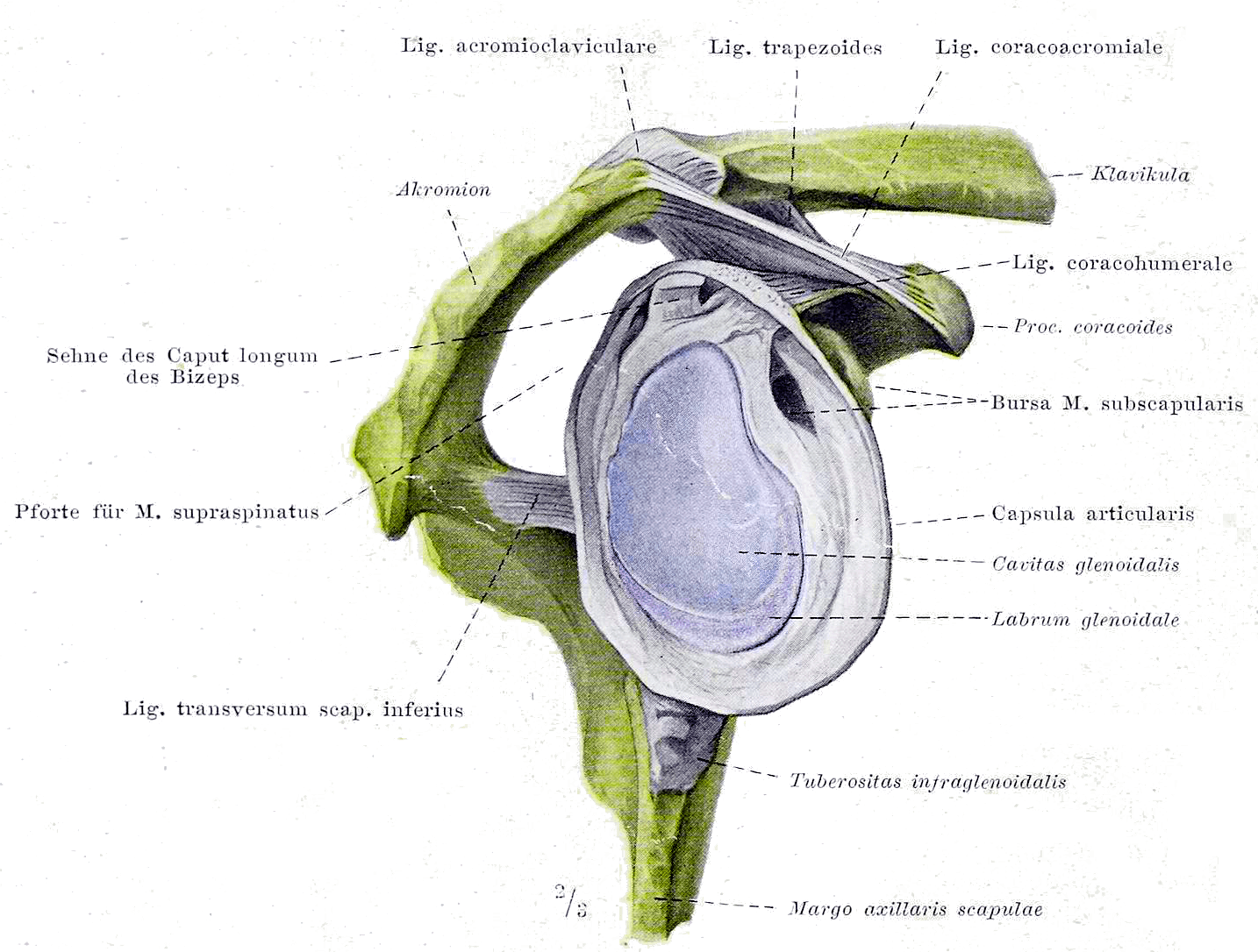
Braus, Hermann, Braus 1921 147, marked as public domain, details on Wikimedia Commons
Treating Subacromial Impingement Syndrome – Subacromial Decompression, Yes or No?
Subacromial decompression (acromioplasty) is a widely used minimally invasive surgical technique for shoulder impingement syndrome.
The procedure usually takes an hour under general anesthesia. Surgical instruments are inserted through tiny incisions (typically 3 incisions with about 2 stitches each). During the surgery, bone is shaved from the underside of the acromion and the bursa may be removed (bursectomy). This is meant to create more space in the subacromial gliding mechanism and resolve the impingement. Additionally, the surgeon repairs any damage to the tendons.
As mentioned in our article Meniscus Injuries in Arthritic Knees: Arthroscopy Yes or No?, we at BodyLab | Osteopathy and Physical Therapy | Rehabilitation and Training | Zurich always ask ourselves what real value surgery brings to the patient.
A recently published review [1] analyzed 9 studies with a total of 1014 patients aged 43 to 57, evaluating pain, joint function, Global Perceived Effect (GPE), and quality of life at 6 and 12 months post-surgery. The control groups in these studies consisted of active physical therapy (6 studies), placebo surgery (2 studies), laser treatments (1 study), bursectomy alone (1 study), and observation (1 study).
The following results were found for the respective factors:
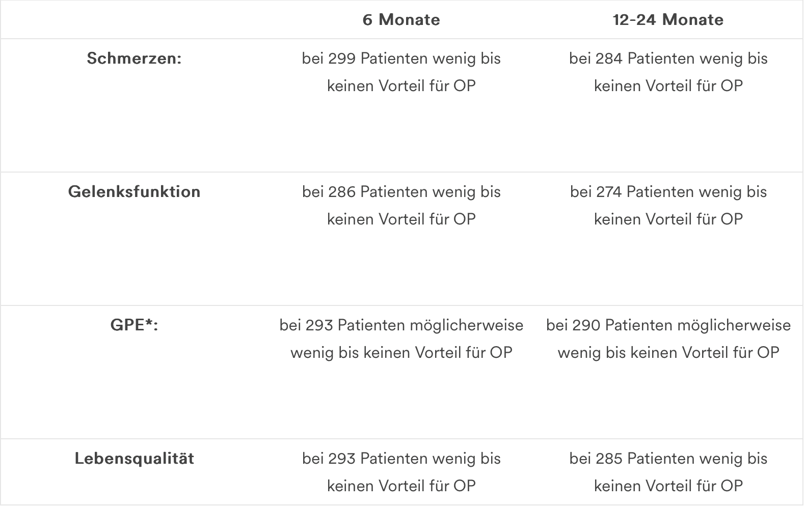
*moderate evidence due to imprecise measurements
It was previously accepted that acromioplasty is a standard procedure for subacromial impingement. However, clear advantages over conservative therapy (exercise/training, physical therapy) have not been definitely proven by clinical studies.
Recent findings show that compared to placebo surgery, there is no significant difference in pain, joint function, or quality of life at 6 to 12-24 months post-surgery. Compared to active physical therapy, there is a small, insignificant difference in pain relief (moderate evidence) and slightly fewer persistent limitations in mobility/joint function (low evidence).
In conclusion, subacromial decompression as a default solution for shoulder impingement should be reconsidered by doctors, healthcare professionals, hospitals, and health insurance providers.
As physical therapists and osteopaths, we highly recommend carefully weighing the pros and cons of surgery and always starting with a conservative approach whenever possible.
Conservative Treatment Options for Shoulder Impingement
The goal of conservative treatment in physical therapy or osteopathy is to restore the biomechanics of the shoulder girdle in subacromial or shoulder impingement syndrome, while stabilizing and strengthening the shoulder. Especially in osteopathy, manual mobilization and manipulation techniques, as well as fascial and soft tissue techniques, are used to release the joints and muscles of the thoracic spine and shoulder girdle to relieve pain and improve mobility.
In physical therapy, once anti-inflammatory measures are taken, the focus shifts to activating the rotator cuff to stabilize and center the glenohumeral joint. Following this, active exercises are used to teach the scapulathoracic rhythm and train the active stabilization of the scapula on the chest wall.
If you need us, we are here for you!
Your BodyLab Team, your team for a free, pain-free shoulder
Osteopathy and Physical Therapy | Rehabilitation and Training
Zurich Altstetten
References
Lähdeoja T, Karjalainen T, Jokihaara J, et al
British Journal of Sports Medicine Published Online First: 15 January 2019. doi: 10.1136/bjsports-2018-100486
Cover Image Credits
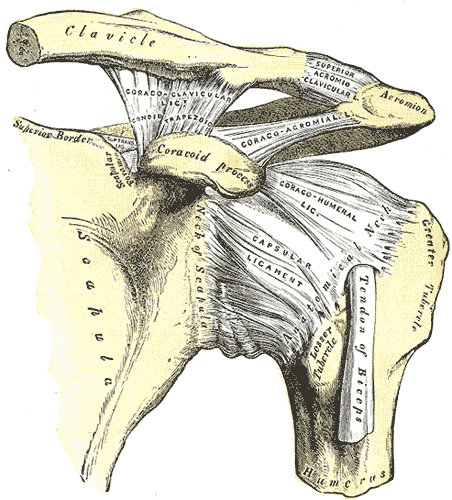
Henry Vandyke Carter creator QS:P170,Q955620 Henry Gray author QS:P170,Q40319, Gray326, marked as public domain, details on Wikimedia Commons