Mindset
Abdominal separation after pregnancy
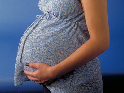
After pregnancy, a visible and palpable gap often forms between your superficial rectus abdominis muscles – known as diastasis recti. Not only can you find this visually bothersome, but it can also affect your entire body. How does diastasis recti impact your body? And are there therapies or exercise programs that can improve your recovery?
Definition
Diastasis recti describes the separation of the two rectus abdominis muscles along the vertical midline of your abdomen (linea alba) [1].
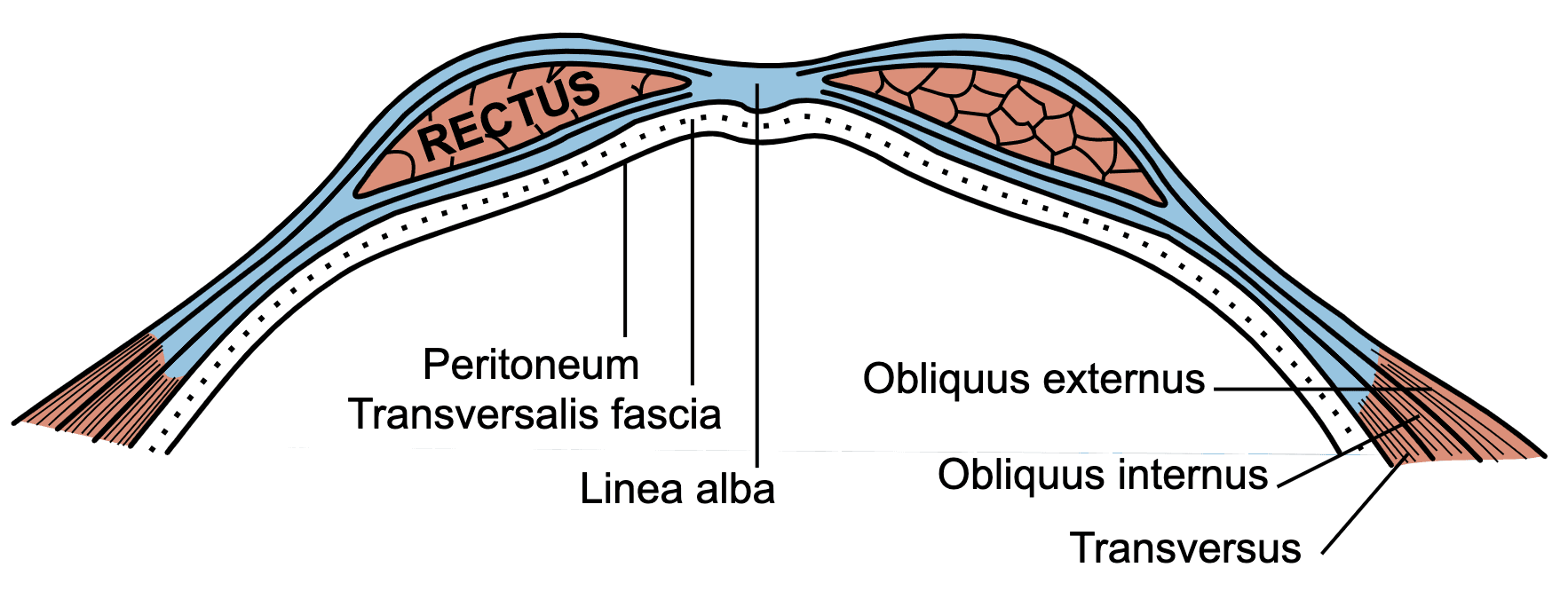
Henry Vandyke Carter creator QS:P170,Q955620 Mysid (SVG) Henry Gray author QS:P50,Q40319, Gray399, marked as public domain, details on Wikimedia Commons
It is diagnosed when a palpable gap of more than two centimeters has developed. Normally, the distance (inter-rectus distance) between the two rectus abdominis muscles is only about 20 mm wide [2]. Diastasis recti can occur in both sexes and in all age groups, but it is most common in pregnant women [3].
Ken Hammond (USDA), PregnantWoman, marked as public domain, details on Wikimedia Commons
During pregnancy, hormonal changes make your abdominal muscles more flexible. The two-bellied rectus abdominis muscle thins out and shifts to the side to create space for your growing uterus. Due to this topographical shift, both rectus abdominis muscles to the right and left of the linea alba lose their optimal line of pull. They move out of their alignment, which is predetermined by their straight fiber direction, and their function becomes compromised. Since the connective tissue of the rectus sheath becomes very thin toward the end of pregnancy due to connective tissue changes, the rectus muscles can easily separate outward. In the early postpartum period, up to the tenth day after birth, a gap of 1–2 finger widths between the muscle bellies is considered physiological. If the distance is wider, it is referred to as diastasis recti; your body loses core support. Diastasis recti then becomes visible as a dome-like protrusion of your internal organs between the separated rectus muscles whenever intra-abdominal pressure increases (e.g., during a crunch or trunk flex). If left untreated, such a diastasis recti can persist for months or even years in some women [4].

Photo by www.localfitness.com.au, Ronnie Coleman 8 x Mr Olympia – 2009 – 5, CC BY-SA 3.0
Effects and Connections
Diastasis recti develops during the course of pregnancy and most commonly becomes noticeable starting in the third trimester. However, it often persists after delivery [4]. In a study by Blaschak & Boissonnault [5], 66% of the 71 tested women had diastasis recti in the third trimester. Directly after birth, the rate was still 53%, and between the fifth and seventh weeks postpartum, 36% of participants still showed a diastasis. This demonstrates that although the incidence of diastasis recti decreases in the postpartum period, it does not spontaneously disappear in all women after giving birth [5]. An existing diastasis recti is often associated with Conditions such as lower back and back pain or incontinence [6]. It is believed that the stabilization of your lower spine can no longer be fully guaranteed. As a result, your overall body posture changes from your feet up to your head position; your rib cage (thorax) and your breathing patterns can also be affected. This dysfunctional breathing and altered posture then affect your pelvic floor and your abdominal and pelvic organs [4]. However, the current scientific literature has not sufficiently clarified the extent to which diastasis recti influences these mentioned Conditions [6]. In their systematic review, Benjamin et al. [6] investigated whether diastasis recti influences musculoskeletal dysfunctions, pain, and quality of life. They identified a link between diastasis recti and the occurrence of pelvic organ prolapse. They also found that diastasis recti can affect health-related quality of life, abdominal muscle strength, and the intensity of lower back pain. However, they found no significant link between diastasis recti and the occurrence of incontinence or pelvic pain [6]. To what extent diastasis recti is linked to other Conditions still needs further investigation before definitive conclusions can be drawn.
Rehabilitation and Training
When dealing with diastasis recti, Physiotherapy and osteopathy are recommended as conservative treatment options. However, there is currently no universally accepted exercise protocol or official treatment guidelines [7]. There are countless different treatment approaches and therapies, but very little research in this area. To date, experts are divided on whether the rectus diastasis should be manually approximated during training or not. The impact of the deep abdominal muscles (transversus abdominis) on diastasis recti is also not yet fully understood. However, it is believed that activating the deep abdominal muscles pulls the diastasis recti further apart, which could be counterproductive. A study by Thabet et al. [8] showed that an exercise program combining passive approximation of the linea alba, pelvic floor activation, breathing exercises, and planks led to a greater reduction in inter-rectus distance than classic abdominal training alone. In a study by Gluppe et al. [9], however, a group of women who completed a weekly exercise program for their abdominal and pelvic floor muscles was compared with a group that did not perform any exercises. No differences could be observed between the two groups even after several months. The scientific evidence is therefore very contradictory. Although there are some promising results, it has not yet been proven that targeted exercise programs can reduce diastasis recti. However, it should be noted that inter-rectus distance has mostly been the only progress metric studied so far. Recent findings show that not only the distance between the muscle bellies, but also the tension of the linea alba is crucial. This tension ensures that your abdominal contents are well supported and that your abdominal muscles can function optimally [10]. It is therefore possible that while physiotherapeutic exercises might not alter the distance itself, they positively influence tissue tension and thus crucial functions. In addition to conservative treatment options, diastasis recti can also be treated surgically. However, this is only medically indicated if an abdominal hernia has developed alongside the diastasis recti [11] or if you have achieved no success with conservative therapy [7].
Conclusion
So far, there is unfortunately little scientific research in this area, and meaningful, standardized testing procedures to objectively monitor the course of diastasis recti are lacking. There are many treatment approaches that are not yet sufficiently evidence-based. However, based on the fundamental principles of exercise science, training your abdominal muscles should be performed in different starting positions and with increasing levels of difficulty, addressing the specific demands and positions that you still find challenging. In this way, personalized and function-oriented therapy with specific exercises can improve your health-related quality of life and reduce restrictions in your daily routine.
Our physiotherapists and osteopaths will gladly support you during your postnatal recovery after pregnancy with all their expertise.
Whenever you need us, we are here for you!
Your BodyLab team, by your side for your postpartum recovery
Osteopathy and Physiotherapy | Rehabilitation and Training
Zurich Altstetten
Find out more on our page pregnancy.
Bibliography
D. R. Benjamin, A. T. M. van de Water, and C. L. Peiris
Physiother. (United Kingdom), vol. 100, no. 1, pp. 1–8, 2014, doi: 10.1016/j.physio.2013.08.005.
[2] The normal width of the linea alba in nulliparous women
G. Beer, A. Schuster, and B. Seifert
Clin Anat., vol. 22, no. 6, pp. 706–711, 2009, doi: 10.1002.
[3] Management Strategies for Diastasis Recti
M. Y. Nahabedian
Semin. Plast. Surg., vol. 32, no. 3, pp. 147–153, 2018, doi: 10.1055/s-0038-1661380.
[4] Nach der Geburt Wochenbett und Rückbildung
A. Heller
1st ed. D-70469 Stuttgard: Georg Thieme Verlag, 2002.
[5] Incidence of diastasis recti abdominis during the childbearing year
M. J. Blaschak and J. S. Boissonnault
Phys. Ther. 1988 Jul;68(7):1082-6. doi: 10.1093/ptj/68.7.1082.
D. R. Benjamin, H. C. Frawley, N. Shields, A. T. M. van de Water, and N. F. Taylor
Physiotheerapy. 2019 Mar;105(1):24-34. doi: 10.1016/j.physio.2018.07.002. Epub 2018 Jul 24.
[7] Diastasis recti abdominis – A review of treatment methods
A. Michalska, W. Rokita, D. Wolder, J. Pogorzelska, and K. Kaczmarczyk
Ginekol. Pol., vol. 89, no. 2, pp. 97–101, 2018, doi: 10.5603/GP.a2018.0016.
A. A. Thabet and M. A. Alshehri
J. Musculoskelet. Neuronal Interact., vol. 19, no. 1, pp. 62–68, 2019.
S. L. Gluppe, G. Hilde, M. Tennfjord, and E. Engh
Annu. Rev. CyberTherapy Telemed., vol. 11, no. 4, p. 63, 2013, doi: 10.1097/01.numa.0000435373.80608.40.
D. Lee and P. W. Hodges
J. Orthop. Sports Phys. Ther., vol. 46, no. 7, pp. 580–589, 2016, doi: 10.2519/jospt.2016.6536.
W. Reinpold et al.
Front. Surg., vol. 6, no. January, pp. 1–6, 2019, doi: 10.3389/fsurg.2019.00001.
Cover Image Credit
Ken Hammond (USDA), PregnantWoman, marked as public domain, details on Wikimedia Commons