Knee
Anterior Cruciate Ligament - Rehab and Prevention

Blog Series - Anterior Cruciate Ligament (ACL) No. 2
Rehab and Prevention
Incidence
As we already highlighted in our first blog on cruciate ligament and knee injuries , injuries to this joint and its surrounding structures are extremely common, especially regarding the anterior cruciate ligament. We see this daily at our BodyLab Osteopathy and Physiotherapy clinic in Zurich, where we consistently treat patients with knee injuries. According to Swiss accident statistics, there was an average of around 50,000 occupational and non-occupational accidents per year between 2012 and 2016 in which the knee was injured in the form of a dislocation, sprain, or strain. The costs for these knee injuries averaged more than 500 million Swiss francs annually over the same period [1]. In a ten-year study, Majewski, Habelt, and Steinbrück categorized all registered sports-related knee injuries into different types [2]. A total of 7,769 knee injuries were recorded during this period, of which 3,482 involved either the cruciate ligaments, collateral ligaments or the menisci; these were grouped together as internal knee injuries. Almost half of all internal knee injuries affected the anterior cruciate ligament (ACL) alone [2].
Costs and Consequences of an Anterior Cruciate Ligament Tear
These injuries don't just cost the healthcare system millions of francs every year; acute knee injuries can also lead to degenerative diseases such as chondropathy or osteoarthritis due to biochemical processes in the joint. These secondary issues can lead to increased limitations in daily life and sports as you age, racking up even higher costs than the original injury itself [3]. Myklebust and Bahr even showed that 50-100% of women who suffer an ACL tear will show radiological signs of knee osteoarthritis in the following 10-20 years [4]. In their review, the expert group of the German Knee Society (DKG) also highlights the fact that 7-24% of patients with a reconstructed ACL go on to suffer a tear in the uninjured knee [5]. For young athletes (<26) competing in high-risk sports (e.g., soccer, volleyball, handball), almost one in four athletes will tear their ACL a second time after returning to their sport. This happens most frequently during the early Return-to-Play (RTP) phase, right after rehabilitation is completed [6]. Compared to uninjured healthy peers, the risk of an ACL injury in patients with a reconstruction increases by 30 to 40 times [6].
Furthermore, post-pubescent women playing the same sports as men face a 2- to 10-fold higher risk of ACL injury right from the start due to various factors like hormonal balance and neuromuscular control [7].
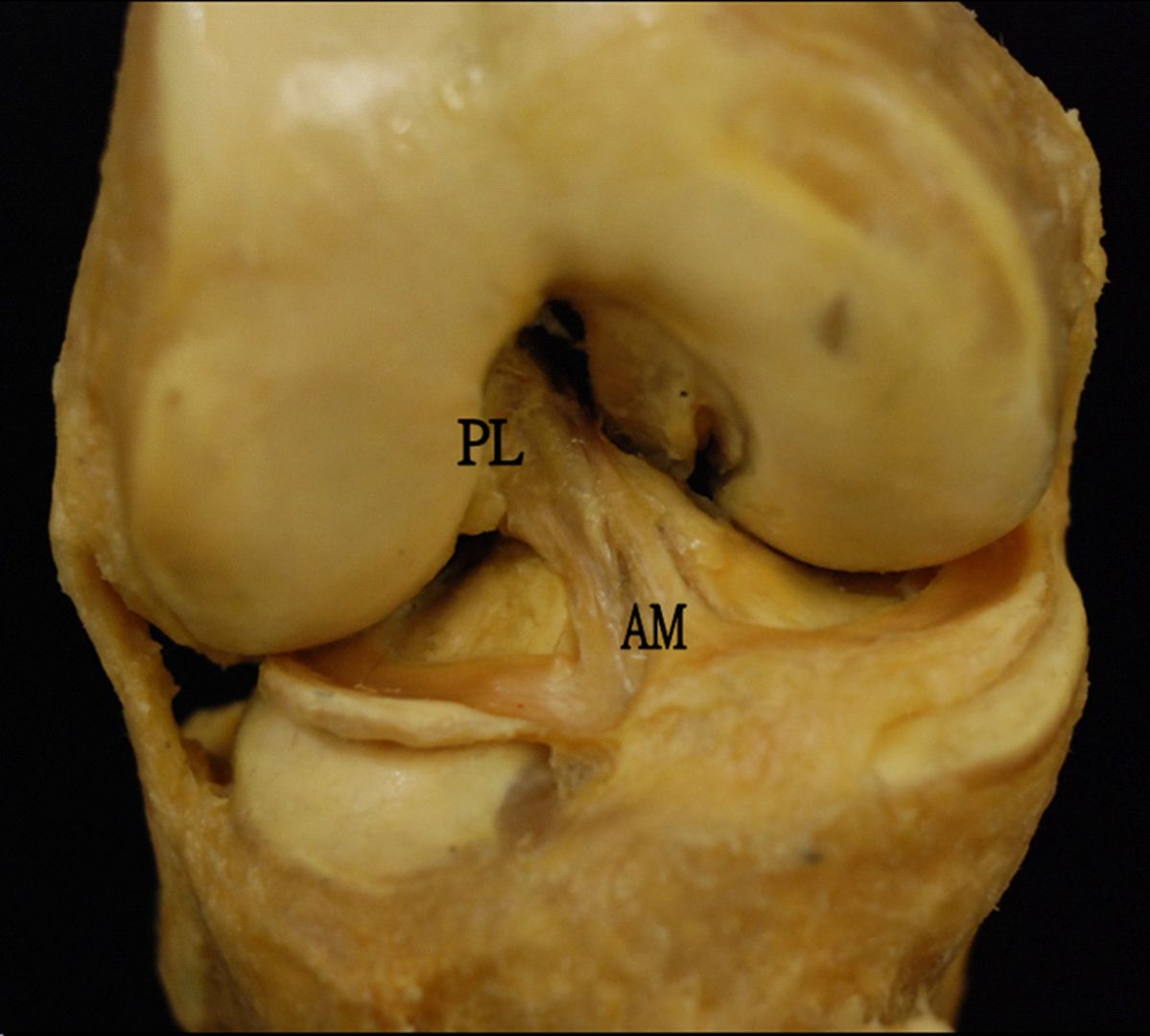
You can find out more about the anatomy and function of the cruciate ligaments in our first blog.
Risk Factors
Scientific literature describes various risk factors that can predispose you to an ACL tear. These are divided into two categories. On one hand, there are extrinsic risk factors like weather conditions, clothing, gear, or the playing surface. On the other hand, we have intrinsic risk factors. These intrinsic factors can be anatomical, physiological, neuromuscular, genetic, or biomechanical in nature [12].
Anatomical risk factors primarily refer to the length, width, and volume of the cruciate ligaments. Additionally, differences in the angle of the cruciate ligaments also influence the risk of an ACL injury.
According to the study by Quatman et al., the combination of anterior tibial translation and internal tibial rotation, as well as abduction and anterior tibial translation, led to a 3.9-fold and 4.6-fold load on the ACL, respectively, compared to normal landing conditions [18].
Landing in a knee-valgus position further increases the load on the ACL because the ground reaction force is elevated in this position. This is a major reason why women face an increased risk of ACL tears. In non-contact ACL tears, the body's center of gravity is often behind the affected knee. Contracting the quadriceps femoris muscle pulls the tibia forward. This puts the ACL under high tension. To avoid losing balance, the hip flexes, which forces the quadriceps femoris to contract even further. Due to an unfavorable lever arm, the hamstrings can no longer adequately support the ACL from behind. As a result, the ACL is subjected to extreme stress, and the risk of a tear spikes in this position [11, 18].
As neuromuscular risk factors, Myers et al. describe reduced hamstring strength relative to quadriceps strength [19]. Pfeifer et al. also succeeded in finding that the hamstrings fatigue faster, which increases the imbalance between the quadriceps and hamstring muscles [12]. This also leads to an elevated risk of ACL tears.
Additionally, research clearly shows that biomechanical risk factors—such as poor landing technique and knee positioning—can be corrected through neuromuscular training programs, making them a powerful strategy for injury prevention (Hewett, Myer, and Ford [17]).
Nessler, Denney, and Sampley concluded from their study that the valgus position combined with limited neuromuscular control of the knee poses the greatest threat for an ACL injury [13]. These risk factors can be proactively addressed with targeted training. To date, neuromuscular training is considered the only effective method to reduce the incidence of ACL tears [7].
Injury Mechanisms
To understand ACL lesions more specifically, they are categorized into different injury mechanisms. A "non-contact injury" refers to an injury where no physical contact or external force from another player caused the damage. Non-contact injuries account for about 72-95% of all ACL injuries [14, 15]. Conversely, a "contact injury" occurs when a direct force hits the knee, causing the injury [16]. ACL injuries involving another player but without direct force applied to the knee are described by researchers as "non-contact injuries with perturbation" [17]. In handball, a study by Olsen et al. identified two movement patterns primarily responsible for non-contact ACL injuries [16]. Both situations—single-leg landing after a jump shot and rapid cutting—result in a valgus knee position with slight flexion and tibia rotation.
We dive deeper into other common injury mechanisms, such as skiing accidents or soccer injuries, right here in our first blog.
Surgery vs. Conservative Treatment
Although researchers are intensively searching for the ultimate treatment options for ACL tears, there is a lack of high-quality studies on managing this injury. Currently, surgical interventions are often preferred for athletes over conservative therapy, especially when patients have accompanying meniscus or medial collateral ligament injuries, or when there is a pronounced feeling of instability [8, 9]. Rehabilitation after an ACL tear followed by surgery takes several months. The literature suggests different timelines depending on the sport, ranging from about 6 to 13 months. Currently, however, there is not enough data to determine whether athletes from different sports actually require different rehabilitation periods before returning to play. Despite this, 83% of elite athletes successfully return to their sport after ACL reconstruction [10].
The Bottom Line
Knee injuries, and anterior cruciate ligament tears in particular (whether they involve other joint structures or not), are among the most common everyday and sports-related musculoskeletal injuries.
It is still unclear whether surgery is absolutely necessary, and this decision must be carefully weighed on a case-by-case basis.
To date, neuromuscular training is considered the only highly effective method to reduce the incidence of ACL tears.
A full return to daily life and sports is absolutely achievable in most cases. However, the real danger lies in the Return-to-Play (RTP) phase, where many athletes end up injuring themselves a second time.
In our next blog about cruciate ligaments, we will take a closer look at your treatment options.
If you need us, we are here to support you every step of the way!
Your BodyLab Team, your experts for prevention and a stable knee
Osteopathy and Physiotherapy | Rehabilitation and Training
Zurich Altstetten
References
[1] Accident Statistics UVG 2018
KSUV
Coord. for Statistics of Accident Insurance UVG, vol. 1, no. 1, pp. 1–64, 2018.
[2] Epidemiology of athletic knee injuries: A 10-year study
M. Majewski, S. Habelt, K. Steinbrück
Knee, vol. 13, no. 3, pp. 184–188, 2006.
L. S. Lohmander, H. Roos, L. Dahlberg, L. A. Hoerrner, M. W. Lark
J. Orthop. Res., vol. 12, no. 1, pp. 21–28, 1994.
[4] Return to play guidelines after anterior cruciate ligament surgery
G. Myklebust, R. Bahr
Br. J. Sports Med., vol. 39, no. 3, pp. 127–131, 2005.
[5] Returning to sports after ACL reconstruction
W. Petersen et al.
Dtsch. Arztebl., vol. 5, no. 3, pp. 166–176, 2016.
A. J. Wiggins, R. K. Grandhi, D. K. Schneider, D. Stanfiled, K. E. Webster, G. D. Myer
Am. J. Sports Med., vol. 44, no. 7, pp. 1861–1876, 2016.
T. E. Hewett, G. D. Myer, K. R. Ford, M. V. Paterno, C. E. Quatman
J. Orthop. Res., vol. 34, no. 11, pp. 1843–1855, 2016.
M. Krause, K. H. Frosch, F. Freudenthaler, A. Achtnich, W. Petersen, and R. Akoto
Dtsch. Arztebl. Int., vol. 115, no. 51–52, pp. 855–862, 2018.
[9] A Systematic Summary of Systematic Reviews on the Topic of the Anterior Cruciate Ligament
M. J. Anderson, W. M. Browning, C. E. Urband, M. A. Kluczynski, and L. J. Bisson
Orthop. J. Sport. Med., vol. 4, no. 3, pp. 1–23, 2016.
C. C. H. Lai, C. L. Ardern, J. A. Feller, and K. E. Webster
Br. J. Sports Med., vol. 52, no. 2, pp. 128–138, 2018.
J. Mehl et al.
Arch. Orthop. Trauma Surg., vol. 138, no. 1, pp. 51–61, 2018.
[12] Risk Factors Associated With Non-Contact Anterior Cruciate Ligament Injury: a Systematic Review
C. E. Pfeifer, P. F. Beattie, R. S. Sacko, and A. Hand
Int. J. Sports Phys. Ther., vol. 13, no. 4, pp. 575–587, 2018.
[13] ACL Injury Prevention: What Does Research Tell Us?
T. Nessler, L. Denney, and J. Sampley
Curr. Rev. Musculoskelet. Med., vol. 10, no. 3, pp. 281–288, 2017.
[14] PROMETHEUS General Anatomy and Musculoskeletal System
E. Schulte, U. Schumacher, and M. Schünke
Stuttgart: Thieme, 2018.
[15] Knee Anatomy
N. N., KLINIK am RING, 2019.
[Online]. Available: https://klinikam-ring.de/orthopaedie/erkrankungen/kniegelenk/anatomie-kniegelenk/. [Accessed: 25-Feb-2019].
O. E. Olsen, G. Myklebust, L. Engebretsen, and R. Bahr
Am. J. Sports Med., vol. 32, no. 4, pp. 1002–1012, 2004.
[17] Anterior cruciate ligament injuries in female athletes: Part 1, mechanisms and risk factors
T. E. Hewett, G. D. Myer, and K. R. Ford
Am. J. Sports Med., vol. 34, no. 2, pp. 299–311, 2006.
C. E. Quatman et al.
ACL Inj. Female Athl., vol. 39, no. 8, pp. 1706–1713, 2011.
G. D. Myer, K. R. Ford, K. D. Barber Foss, C. Liu, T. G. Nick, and T. E. Hewett
Clin. J. Sport Med., vol. 19, no. 1, pp. 3–8, 2009.
Cover Image Credit
Mak-Ham Lam, Daniel TP Fong, Patrick SH Yung, Eric PY Ho, Wood-Yee Chan and Kai-Ming Chan, ACLI 18, CC BY-SA 2.0