Foot & Ankle
Acute Supination Injury - Ligament Injuries of the Foot
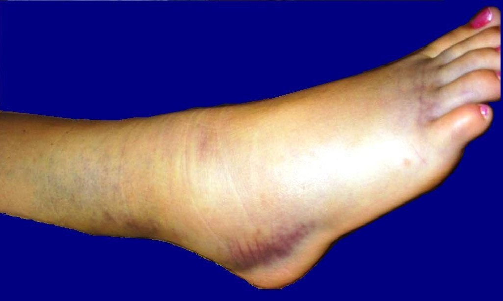
Inversion sprain, sprain, ligament injuries, or torn ligaments of the foot
Ligament injuries of the ankle (inversion traumas, ankle sprain, torn ligament) are very common and frequent injuries, whether in everyday life from a simple misstep or, even more often, during a wide variety of leisure and sports activities (see BFU accident statistics): An ankle injury is one of the most common musculoskeletal injuries of the human locomotor system worldwide.
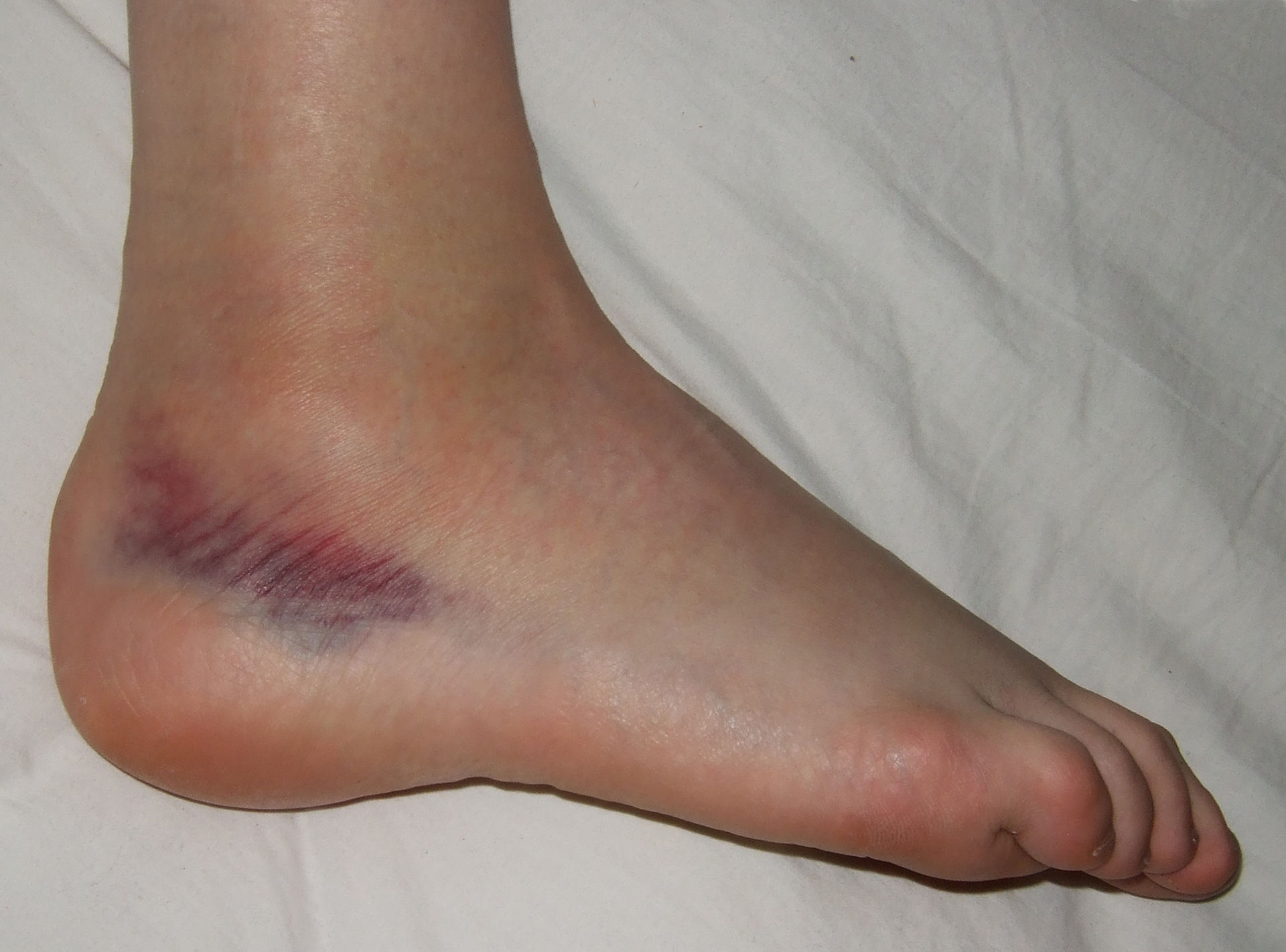
Boldie, Sprained foot, marked as public domain, details on Wikimedia Commons
Nevertheless, at BodyLab osteopathy and Physiotherapy, Rehabilitation and Training in Zurich, we see completely different approaches and medical care depending on the hospital or doctor: from casts and braces with or without walking aids to tape bandages or no immobilizing support at all and purely a referral to physiotherapy—especially among sports doctors…
Due to the high number of injuries, numerous scientific publications and studies address this topic. And we at BodyLab have also done some literature research.
As already mentioned in our blog post on cruciate ligament injuries, various countries have their own committees / medical boards that develop and publish guidelines for such common injuries based on the latest scientific evidence. Guidelines are recommendations meant to help your therapist assess whether your injury's wound healing process is progressing normally or not, and they examine and recommend appropriate therapeutic and treatment interventions. They provide a framework and serve as a reference.
We find the Dutch «KNGF-Guideline for physical therapy in patients with acute ankle sprain» to be an important and valuable paper: in the Netherlands, patients can go directly to a physiotherapist (without a doctor's referral), which means a physiotherapist needs to know what must be done and when! They have to be able to rely on their differential diagnostics—and master them.
Anatomy and function of foot ligaments – capsule
Your ankle joint provides the connection and therefore the transfer of force from your body/lower leg to your foot/ground. It consists of two joint sections—the upper ankle joint, formed by the connection of the fibula and tibia (malleoli) with the talus, and the lower ankle joint where the talus meets the calcaneus.
Most of the stability in your ankle is achieved through the arrangement of the bones and the capsular structure of the numerous ligaments. The muscles around your ankle originate in the lower leg and, with the exception of the Achilles tendon, are redirected via retinacula (ligamentous structures) or bony grooves. Consequently, the muscles do not attach directly to the ankle joint.
The joint is stabilized medially and laterally by ligaments. On the medial side, the broad deltoid ligament fans out from the inner ankle to the talus, the navicular bone (tarsal bone), and the calcaneus (heel bone). Anatomically, the deltoid ligament is divided into four parts. The main function of the deltoid ligament is to stabilize against inward rolling (valgus stress) and to inhibit eversion. It is one of the strongest ligaments in your body.
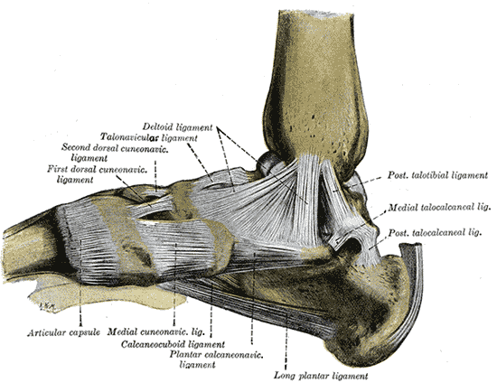
Henry Vandyke Carter creator QS:P170,Q955620 Henry Gray creator QS:P170,Q40319, Gray354, marked as public domain, details on Wikimedia Commons
On the outside, three different ligaments make up the lateral collateral ligament complex—the calcaneofibular ligament and the anterior and posterior talofibular ligaments. They each run from the outer ankle to the calcaneus or parts of the talus, respectively.
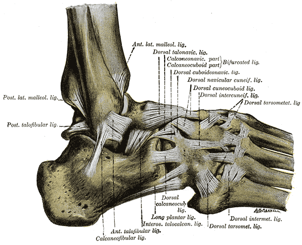
Henry Vandyke Carter creator QS:P170,Q955620 Henry Graycreator QS:P170,Q40319, Gray355, marked as public domain, details on Wikimedia Commons
Just from the structural differences between the medial ligament and the lateral ligaments (width and arrangement), you can tell that the inside of your ankle joint is stabilized much more strongly by passive structures. You can also feel for yourself that your foot can move further inward than outward.
If the "weaker" lateral ligaments are subjected to excessive tension when you take a misstep and twist your foot, these outer ligaments can easily become overstretched or torn.
Injuries and injury mechanisms
When you twist your ankle and roll your foot inward, excessive movement occurs, which usually affects the anterior talofibular ligament first. Depending on the severity and the force involved, this ligament can be overstretched or even torn. Further capsular injuries can occur (since the ligaments are mostly fused with the capsule), as well as additional ligament tears in the foot (e.g., the bifurcate ligament) or even syndesmosis injuries (the connection between the tibia and fibula). Avulsion fractures or bone fractures can also occur.
Due to the rectangular anatomical shape of the talus, impaction injuries of the articular cartilage in the tibiofibular mortise of the upper ankle joint frequently occur, along with bone bruises. When the talus tilts too sharply, it creates high, pinpoint pressure in the upper ankle joint, causing you to feel Conditions and pain on the inside of the joint as well.
Screening process
The goal is to identify your problem and the injured structures, as well as to recognize red flags (severe injuries such as fractures). Do your symptoms match the known clinical signs of an ankle sprain?
Is it an acute sprain caused by an accident? Or does the problem stem from chronic or functional instability? To detect or rule out a fracture after an acute accident, the Ottawa Ankle Rules are helpful. These have nearly 100% sensitivity (the sensitivity of a diagnostic test indicates how often the condition is correctly identified in patients who actually have it), but only moderate (25–50%) specificity (which indicates the proportion of healthy individuals correctly identified as not having the condition).
The Ottawa Ankle Rules state:
If you cannot walk four steps immediately after the injury, or
you experience increased local bone tenderness along the posterior edges or tips of the malleoli (ankle bones) at four palpation points, or
you experience increased bone tenderness at the navicular bone or the base of the fifth metatarsal,
a medical consultation with an X-ray is indicated.
Diagnostic process
The goal of the physiotherapy examination and diagnosis is to use medical and other information to determine whether Physiotherapy can influence and modify the triggering and limiting factors that led to your health problem, as confirmed by your physiotherapist.
The diagnostic process should lead to a clear physiotherapy diagnosis!
Your therapist examines and determines the recency and severity of the injury to your capsular-ligamentous complex based on your medical history and examination. The functional examination includes active tests (active range of motion, whether you can put weight on your foot) as well as passive and palpation tests. With these, a differential diagnosis is made to evaluate which structures are injured (muscle, tendon, cartilage, or capsular-ligamentous structures).
Afterward, your therapist should also be clear on which healing phase your tissue is in and whether a normal or abnormal healing process is taking place. This allows for a prognosis and provides insights into physiotherapy treatment options.
Therapy
We recommend starting physiotherapy measures as soon as possible (within 5 days to a week).
Therapy varies depending on how recent the injury is, the condition (wound healing phase) of your tissue, and your specific Conditions.
In the acute phase (0–3 days after the injury), pain-relieving and swelling-reducing techniques are indicated. Depending on your pain, partial weight-bearing and elevating your injured foot make sense during this time! A compression bandage can also be considered in this early stage if there is significant swelling. Nevertheless, your foot and toes should be moved actively and pain-free as much as possible.
A very important part of every treatment is also patient education and guidance!
Depending on the severity and stage of healing, the next steps involve gradually restoring functionality and activity, increasing intensity based on your pain level. You begin with functional exercises aimed at restoring your normal range of motion, promoting stability and coordination in your foot, and helping you return to a normal walking pattern.
Over time, exercises are introduced that build foot strength and improve your proprioception.
As we explained in our blog post on wound healing, there is no faster healing than optimal healing! At BodyLab, we have the expertise to provide you with optimal support and guidance for injuries to your locomotor and musculoskeletal system. We are here to relieve your pain and to identify or prevent delays and complications (such as infections or long-term deficits) early on—for your optimal, fastest, and best possible recovery.
Whenever you need us, we are here for you!
Your BodyLab team, your specialists after a twisted ankle, helping you get back on solid ground
Osteopathy and Physiotherapy | Rehabilitation and Training
Zurich Altstetten
Find out more on our page about torn ankle ligaments.
References / Guidelines
KNGF-Guideline for Physical Therapy in patients with acute ankle sprain
Supplement to the Dutch Journal of Physical Therapy, Volume 116 / Issue 5 / 2006
KNGF-Flowchart for Physical Therapy in patients with acute ankle sprain / functional instability
Cover image credits
Harrygouvas, ANKLE SPRAIN 02b, CC BY-SA 3.0
Harrygouvas